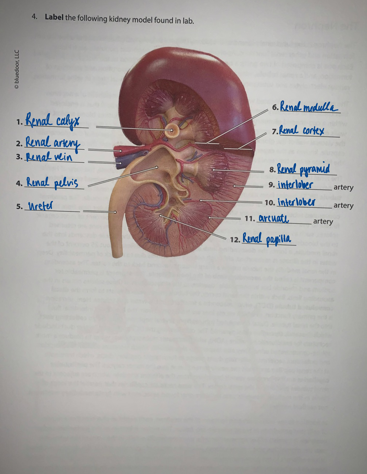
45 picture of the kidney with labels
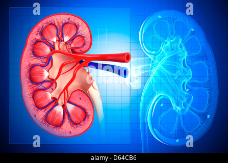
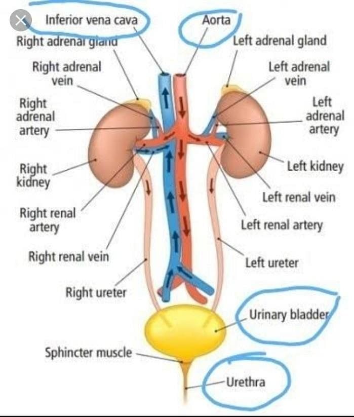
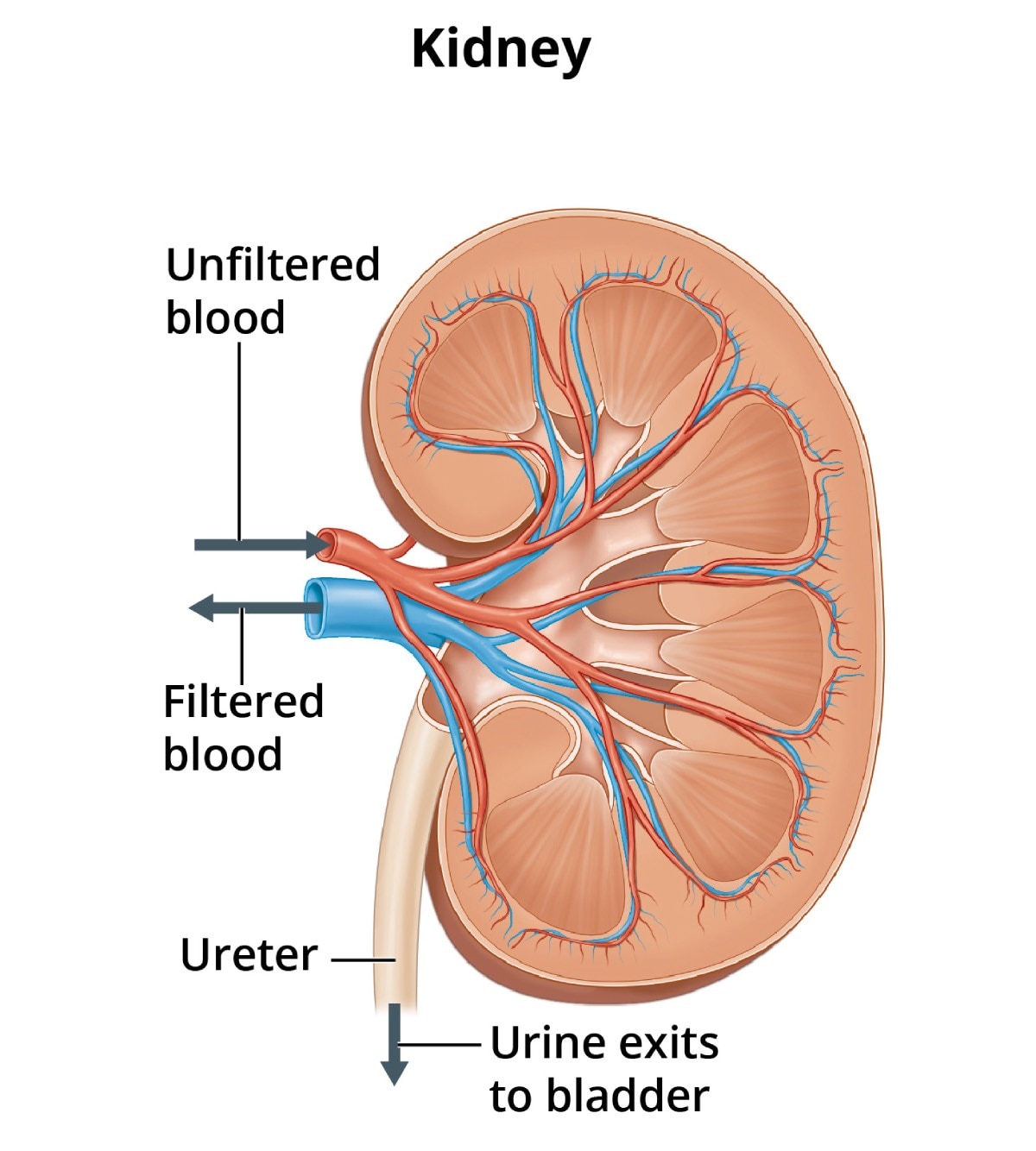
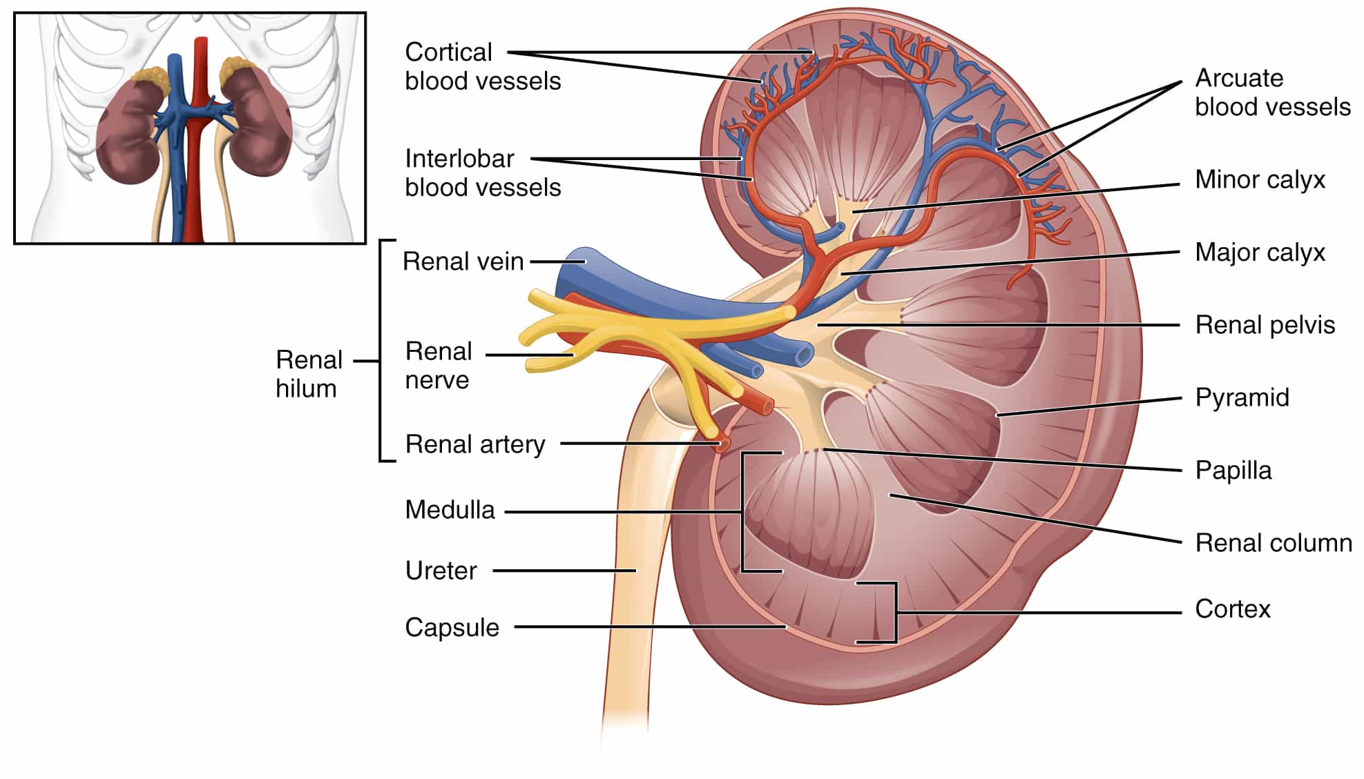
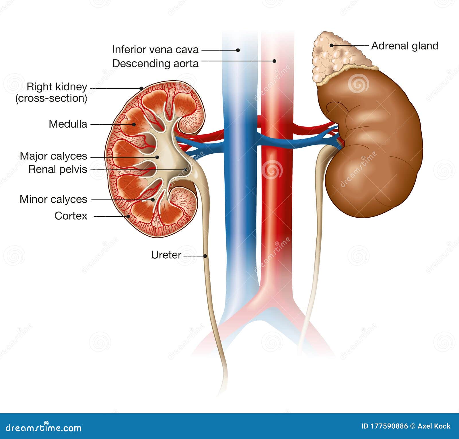
Urinary System - Label the Kidney and Nephron - The Biology Corner Students drag labels to the structures on the slide. Also, the diagram shows the relationship between the aorta, vena cava, and the renal vessels. While these aren't part of the urinary system, they are important in the physiology of the kidney. On the second slide, viewers see a close-up of a kidney that's been cut to show the internal structures. Labeled Diagram Of The Digestive System Stock Photos, Pictures ... Labeled medical diagram with structure and location. Cross section scheme with diverticula infected or inflamed and diverticulosis. Kidney Anatomy Labeled, Cross Section View on White Computer generated image of kidney cross section showing the kidney interior with renal arteries and veins, with anatomy labels on a white background.
Urinary System With Labels , 5 Urinary System Pictures In Organ ... Jun 26, 2019 - urinary system Biological Science Picture Directory - Pulpbits.net. ... Subjects. Science. Visit. Save. Article from . pulpbits.net. urinary system. Urinary System With Labels , 5 Urinary System Pictures In Organ Category. Piyush Razz . Brain Diagram. Chemo Brain. Basal Cell Carcinoma ... Kidney Failure Symptoms. Causes Of Kidney ...

Picture of the kidney with labels
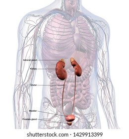
Kidneys: Anatomy, Location, and Function - Verywell Health Anatomy. Each person has two kidneys. The kidneys are located on either side of the spine, with the top of each kidney beginning around the 11th or 12th rib space. The kidneys are sandwiched between the diaphragm and the intestines, closer to the back side of the abdomen. Roughly the size of a closed fist, each kidney measures about 10 to 12 ... Kidney Anatomy Labeling Diagram | Quizlet Start studying Kidney Anatomy Labeling. Learn vocabulary, terms, and more with flashcards, games, and other study tools. Solved Activity 4 - Draw a picture of the kidney and label - Chegg Transcribed image text: Activity 4 - Draw a picture of the kidney and label it with the names of following vessels! The artery and veins that travel through the renal sinus The artery and veins that travel through the renal columns The artery and vein that arch over the base of the renal pyramid between the cortex and the medulla The blood vessels located in the renal cortex . .
Picture of the kidney with labels. Kidney - austincc.edu Kidney. This image is a section that shows almost the entire thickness of the kidney. The outer layer of the kidney--the cortex--is on the top. As you move down through the image you will see part of the kidney medulla. The round structures in this part of the kidney are called renal corpuscles (rc). Each renal corpuscle consists of a ... The 30 Healthiest Foods to Eat Every Day - Real Simple Feb 22, 2022 · Magdalena Niemczyk - ElanArt/Getty Images. The whites offer up protein with minimal calories (and zero fat or cholesterol). Egg yolks get a bad rap, but don't skip them—they are awash with vitamin B12 and vitamin A, and they contain choline, a nutrient that's particularly important for pregnant women. Kidneys: Location, function, anatomy, pictures, and related diseases The kidneys are a pair of bean-shaped organs present in all vertebrates. They remove waste products from the body, maintain balanced electrolyte levels, and regulate blood pressure. The kidneys ... 265 Picture Of Kidneys In Human Body Premium High Res Photos - Getty Images 224 Picture Of Kidneys In Human Body Premium High Res Photos Browse 224 picture of kidneys in human body stock photos and images available, or start a new search to explore more stock photos and images. of 4 NEXT
2,432 Nephron Images, Stock Photos & Vectors | Shutterstock Find Nephron stock images in HD and millions of other royalty-free stock photos, illustrations and vectors in the Shutterstock collection. Thousands of new, high-quality pictures added every day. Tylenol Liver Damage: Signs, Symptoms, Dosages, Overdose ... May 16, 2022 · Tylenol in high doses can permanently damage the liver and lead to coma and death in some cases. Learn about the signs and symptoms of Tylenol-related liver damage, as well as its causes. Discover how Tylenol liver damage is diagnosed, and what treatment options are available. Kidney Structures and Functions Explained (with Picture and Video ... Your kidneys are paired organs found on each side of the back portion of the abdominal cavity. The larger left kidney is located a bit higher than the right kidney. Unlike other organs found in the abdomen, the kidneys are located behind the lining (peritoneum) of the abdominal cavity, thus they are considered retroperitoneal organs. Microscopic Anatomy of the Kidney | Anatomy and Physiology II ... The filtration membrane of the nephron is formed by the fenestrated endothelium of the glomerulus, a basement membrane, and the podocytes of Bowman's capsule. Podocytes have projections that interdigitate to form filtration slits, leaving small gaps between the digits to form a sieve.As blood passes through the glomerulus, 10 to 20 percent of the plasma filters between these sieve-like fingers ...
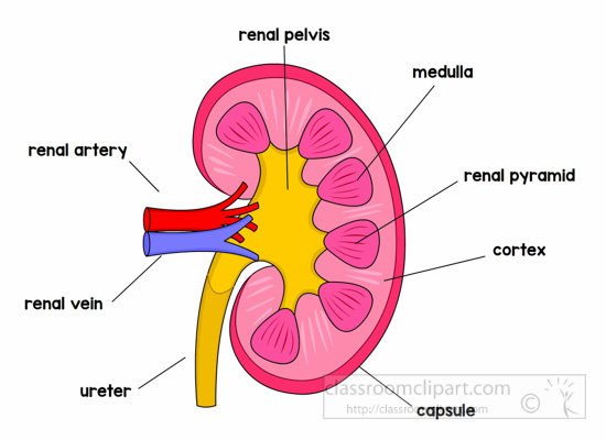
Urinary System Diagram - Kidney, Urinary Tract, Renal ... - SmartDraw Urinary system diagrams are illustrations of the urinary system, also referred to as the renal system. The urinary system, at a high level, contains two kidneys, two ureters, a urethra, and a bladder. The urinary system is located directly below the rib cage. Kidneys - Anatomy Pictures and Information - Innerbody The kidneys are the waste filtering and disposal system of the body. As much as 1/3 of all blood leaving the heart passes into the kidneys to be filtered before flowing to the rest of the body's tissues. While a person could live with only one functioning kidney, our kidneys are vital organs; the loss of both kidneys would lead to a rapid ... Label and Color the Kidney - The Biology Corner Image of the kidney, showing the ureter, renal vessels, cortex, renal pyramids, and renal pelvis. Students label and color the image for practice with anatomy Kidney Images | Free Vectors, Stock Photos & PSD Find & Download Free Graphic Resources for Kidney. 12,000+ Vectors, Stock Photos & PSD files. Free for commercial use High Quality Images
Lesson Worksheet:Kidney Structure | Nagwa
Long COVID or Post-COVID Conditions | CDC Sep 01, 2022 · Multiorgan effects can involve many body systems, including the heart, lung, kidney, skin, and brain. As a result of these effects, people who have had COVID-19 may be more likely to develop new health conditions such as diabetes, heart conditions, or neurological conditions compared with people who have not had COVID-19.

Cenveo - Drawing Kidney in coronal section - English labels ...
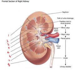
Kidney Anatomy and Function - Health Pages Kidney Anatomy. Renal Capsule - An outer membrane that surrounds the kidney; it is thin but tough and fibrous. Renal Pelvis - Basin-like area that collects urine from the nephrons (the kidney's filtration system), it narrows into the upper end of the ureter. Calyx - The extension of the renal pelvis; they channel urine from the pyramids ...
Human kidney anatomy, cross section with labels Stock Photo ...
Feeding Your Cat: Know the Basics of Feline Nutrition ... This is a crucial point when one considers how common kidney and bladder problems are in the cat. Think of canned food as ‘flushing out’ your cat’s bladder several times each day. Please keep in mind that when your cat starts eating a more appropriately hydrated diet of canned food, his urine output will increase which is a very good ...
Longitudinal Section of a Kidney | ClipArt ETC
Liver And Kidney Pictures Illustrations, Royalty-Free Vector ... - iStock Browse 125 liver and kidney pictures stock illustrations and vector graphics available royalty-free, or start a new search to explore more great stock images and vector art. Newest results Watercolor X-rays of organs Watercolor anatomy collection - X-rays of organs. Heart, lungs, brain, stomach, kidney, liver.
Renal System 1- Structure of the Kidney, Ureters, Bladder and ...
Labeled diagram of the human kidney royalty-free images - Shutterstock 189 labeled diagram of the human kidney stock photos, vectors, and illustrations are available royalty-free. See labeled diagram of the human kidney stock video clips Image type Orientation People Artists Sort by Popular Healthcare and Medical Anatomy Diseases, Viruses, and Disorders kidney organ medicine human body urinary system kidney cancer
199 Labeled Diagram Of The Human Kidney Images, Stock Photos ...
Kidneys (Anatomy): Picture, Function, Conditions, Treatments - WebMD The kidneys are a pair of bean-shaped organs on either side of your spine, below your ribs and behind your belly. Each kidney is about 4 or 5 inches long, roughly the size of a large fist. The...

Kidney Healthy Food Labels Survey
Kidney Labeling Flashcards | Quizlet functional unit of kidney- millions located throughout medulla/cortex border. THIS SET IS OFTEN IN FOLDERS WITH... Kidney vocab and Urinary Disorders. 27 terms. Ms_Maile. Muscle Set arm page 1. 13 terms. Ms_Maile. KIDNEY LABEL. 8 terms. domie4143. Brain structures. 33 terms. Ms_Maile. YOU MIGHT ALSO LIKE... Kidney functions. 14 terms ...

chet_rice_terminolog_2|The Urinary System|Labeling
Illustration Picture of Abdominal Area - Abdomen Structure and Function The abdominal cavity is the part of the body that houses the stomach, liver, pancreas, kidneys, gallbladder, spleen, and the large and small intestines. The diaphragm marks the top of the abdomen and the horizontal line at the level of the top of the pelvis marks the bottom. Connective tissue called the mesentery holds the abdominal organs ...
Draw and label the parts of LS of kidney? - Sarthaks eConnect ...
Picture of the Heart - WebMD Viral infections, kidney failure, and autoimmune conditions are common causes. Pericardial effusion: Fluid between the lining of the heart (pericardium) and the heart itself. Often, this is due to ...

Anatomy of the Kidneys and Renal Blood Vessels | Doctor Stock
CBS Philadelphia - Breaking News, Sports, NEXT Weather ... Phillies' 6-run ninth tops Cardinals in 6-3 wild-card win Game 2 will be played on Saturday night. 13H ago

25 Labeled Diagram Of Kidney Stock Photos, Pictures & Royalty ...
Longitudinal Section of a Kidney | ClipArt ETC - FCIT A longitudinal section of a kidney. Labels: 1, 2, 3, Parts of the Kidney. 4, Pelvis. 5, Ureter. 6, Renal artery. 7, Renal vein. 8, Branches of the latter vessels in the kidney. Keywords kidney Galleries Human Excretory System Source
Human AP Lab 9 - Label the following kidney model - BIOL 2020 ...
1,327 Liver And Kidneys Premium High Res Photos - Getty Images 1,327 Liver And Kidneys Premium High Res Photos Browse 1,327 liver and kidneys stock photos and images available, or start a new search to explore more stock photos and images. of 23 NEXT

BIOL-2311L Internal Structure of Kidney Labeling Diagram ...
Kidneys Picture Image on MedicineNet.com Picture of Kidneys The kidneys are a pair of organs located in the back of the abdomen. Each kidney is about 4 or 5 inches long -- about the size of a fist. The kidneys' function are to filter the blood. All the blood in our bodies passes through the kidneys several times a day.

Kidney Diagram Labeling Quiz - Label The Parts Of The Kidney - Interactive Diagram Online
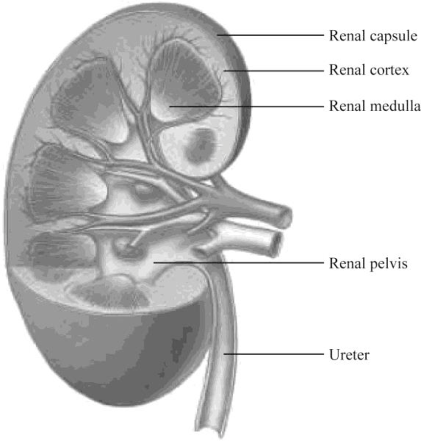
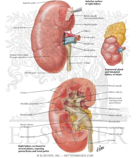
Kidneys: Anatomy, function and internal structure | Kenhub There are 8-18 renal pyramids in each kidney, that on the coronal section look like triangles lined next to each other with their bases directed toward the cortex and apex to the hilum. The apex of the pyramid projects medially toward the renal sinus. This apical projection is called the renal papilla and it opens to the minor calyx.
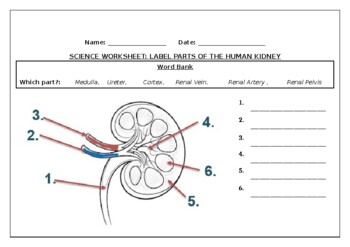
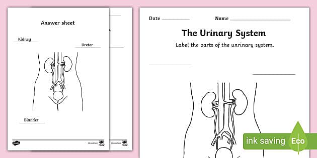
Science worksheets: Label parts of the human kidney
Color and Label the Nephron | Biology diagrams, Nursing school tips ... Practice labeling the nephron with this reinforcement activity. Students can also color the image to identify the major structures of the nephron: glomerulus, bowman's capsule, proximal and distal tubules, loop of henle, collecting duct and capillaries. This was designed to go with a larger unit on how the urinary system and kidneys help the body

Q6 Draw a diagram of the human excretory system and label the ...
Join LiveJournal Password requirements: 6 to 30 characters long; ASCII characters only (characters found on a standard US keyboard); must contain at least 4 different symbols;

Draw the diagram of kidney, recognize and label the following ...
Labeled Diagram of the Human Kidney - Bodytomy Labeled Diagram of the Human Kidney The human kidneys house millions of tiny filtration units called nephrons, which enable our body to retain the vital nutrients, and excrete the unwanted or excess molecules as well as metabolic wastes from the body. In addition, they also play an important role in maintaining the water balance of our body.
Draw the given diagram of LS of kidney and label the class 11 ...
Diagrams showing the structure of kidney cortex and medulla What's inside a kidney? The two main parts of a kidney. The kidneys are made up of two main parts - the outer part, known as the cortex, and the inner part, called the medulla.. The kid who needed a clean-up was wearing a corset (cortex) and a medal (medulla). Note that the medal (medulla) is in the centre of the corset (cortex).. Diagram of cortex and medulla
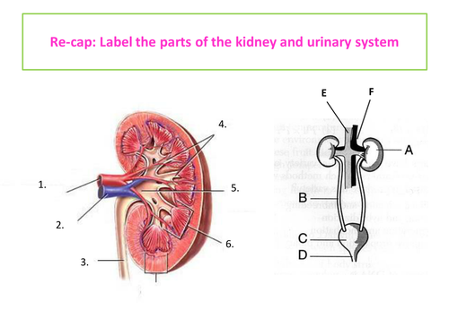
Starter quiz on parts of kidney and urinary system | Teaching ...
Solved Activity 4 - Draw a picture of the kidney and label - Chegg Transcribed image text: Activity 4 - Draw a picture of the kidney and label it with the names of following vessels! The artery and veins that travel through the renal sinus The artery and veins that travel through the renal columns The artery and vein that arch over the base of the renal pyramid between the cortex and the medulla The blood vessels located in the renal cortex . .

Identifying the Different Parts and Structures on the Kidney Diagram
Kidney Anatomy Labeling Diagram | Quizlet Start studying Kidney Anatomy Labeling. Learn vocabulary, terms, and more with flashcards, games, and other study tools.
Draw a diagram of excretory system in human beings and label ...
Kidneys: Anatomy, Location, and Function - Verywell Health Anatomy. Each person has two kidneys. The kidneys are located on either side of the spine, with the top of each kidney beginning around the 11th or 12th rib space. The kidneys are sandwiched between the diaphragm and the intestines, closer to the back side of the abdomen. Roughly the size of a closed fist, each kidney measures about 10 to 12 ...

The kidney and osmoregulation - Ms. Frost A world of biology.....

Sketch a labelled L. S. of human kidney.

Kidney - Wikipedia

Label the kidney diagram Quiz
Your Kidneys & How They Work | NIDDK

0514 Anatomy Of Kidney Medical Images For PowerPoint ...
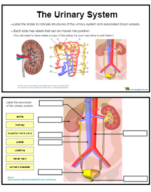
Urinary System – Label the Kidney and Nephron
Anatomy of the Urinary System
Anatomy Clipart - anatomy-kidney-labeled-clipart - Classroom ...

25 Labeled Diagram Of Kidney Stock Photos, Pictures & Royalty ...
To label: The parts of the kidney and nephron. Introduction ...
A Kidney Problem?

AstraZeneca ramps up Farxiga push in new kidney disease label

draw a well labelled diagram of the l s of kidney label any ...
Autoimmune Kidney Disease - Benefits of an Integrative ...
Kidney and Bladder Labelling Worksheet

Kidney Anatomy & Nephron Filtration Diagram photo | Kidney ...

Evaluation of Renal Blood Flow in Chronic Kidney Disease ...
Kidneys Anatomy, Medically Illustration, Labeled Stock Vector ...
Atlas of Human Anatomy - 2nd Edition

Kidneys and ureters isolated in male internal anatomy on ...
Urinary System Anatomy Waste Urinary System Urinary System ...

Activity - Kidney Anatomy_-1072546379 (1).pdf - S T U D E N T ...

Which of the following is the correct labelling of parts of ...
Post a Comment for "45 picture of the kidney with labels"